One of the major challenges in ophthalmic care is to be able to document and diagnose the various problems of binocular vision.
With KMScreen, which is a digital Hess and Harms Screen, you can quickly and easily document various deviations of the extraocular muscles in a 15 ° internal and 25 ° or/and 30° external field *. The test can be complemented by a diplopia test which records the deviation and the torsion between the two images in the nine cardinal positions of gaze. Together with Hess and Harm's program, there is also a program that examines the binocular field of single vision.
KM Screen was developed at the Paediatric and Strabological Department of Eye Clinic, Skåne University Hospital -Sweden-(SUS). It is registered at the Swedish Medical Products Agency as medical equipment authorised to be sold within the EU.
*
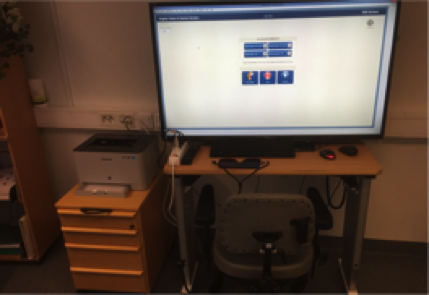
Workspace with 55 inch screen
Hess method: 15 ° / 25 ° Harm method: 15 ° / 25 ° / 30 °
Workspace with 65 inch screen
Hess method: 15 ° / 25 ° / 30 ° Harms method: 15 ° / 25 ° / 30 °
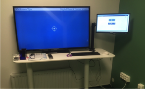
KMScreen is made up of a 55 or 65" inch LED screen and a laptop with pre-installed software. The examiner observe and follow the test results following every click from the patient at the laptops screen.

Each KMScreen unit comes with three types of software. An examining eye deviation according to Hess method and according to Harms method. With the third program we can examine the binocular field of single vision.
The examination using the Harms’ method
The patient keeps his eyes fixated on the center of the screen, while the examiner is turning the patient's head in the nine cardinal gaze directions.
In order to examine the upward gaze, for example, the examiner should turn the patient’s head 25 degrees downwards. Meanwhile, the patient is looking at the fixation object that is always in the center of the screen. The Harms-program can simultaneously test the horizontal-vertical deviation of both eyes and torsion.
Both Hess’ and Harms’ examinations take about 2 to 9 minutes to complete, depending how familiar the patient is with a particular program and the use of computer mouse.
The test distance is 50 cm.
Examination using the Hess’ method
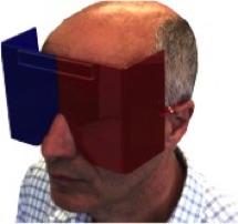
The patient is sitting in front of the TV screen while wearing the red-blue visor glasses and holding his/her head still.
The screen displays two objects: one of them is moving, and the other is stationary. The patient sees the moving object with one eye, and the stationary object with the other. Using a computer mouse, the patient should drag the moving object on the screen towards and over the stationary object. When objects on the screen overlap, the patient is supposed to click on the mouse.
After each click of the mouse, the fixation point is moved to a new position. The patient should continue to hold his/her head perfectly still during the whole examination. Via a secondary display the examiner can observe and follow the test results directly after each click. If necessary, the examiner can go back to the previous point / points again.
Once the patient has finished clicking at all fixation points, the result is shown as a conventional Hess screen diagram. The examiner can then perform an extra diplopia test, which investigates the vertical and horizontal deviation as well as the torsion of the double images in the 9 gaze directions.
The test distance is 50 cm
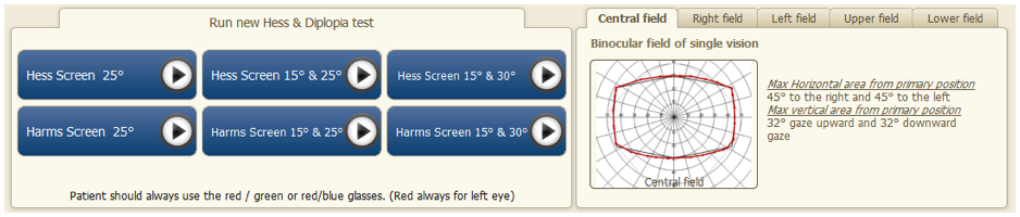
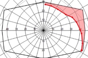
Binocular field of single vision
The binocular field is divided into 24 sectors á 15° each. Tested without the red blue visor glasses.
The patient is sitting in front of the TV screen and holding his/her head still and instructed to follow with the eyes the red ring. The caregiver stand behind the patient and, if necessary, keep the patient's head still with one hand and the wireless mouse against a smooth surface or the thigh with the other hand.
The caregiver moves slowly the red ring from the center to the periphery a sector at a time. When the patient clicks that he/she is seeing double, or we reach the end of this sector without diplopia continue to test whether there is double vision in the next sector that is located next to the one we have tested. The result appears immediately after each click on the secondary screen.
Test results for Hess and Harms are analysed, stored and presented, and can be printed as follows:
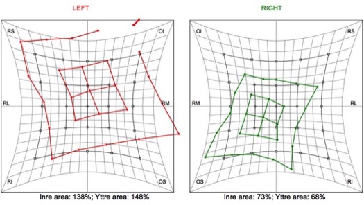
1. As a conventional Hess screen chart where the outer and inner fields (15 ° and 25 °) of both eyes are plotted. The charts of the two eyes are compared for position, size and shape.
2. The outer and inner field area of both eyes is measured in percent. You can compare the two outer fields to see if there is a concomitant or incomitant strabismus. Then, you can compare the outer area with the inner area of each eye to diagnose if there is a mechanical limitation of the muscles in the eye.
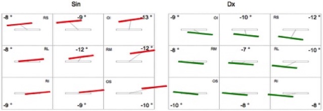
3. Diplopia chart in nine gaze directions where the horizontal and vertical deviation and the torsion of the double images are plotted.
The torsion is given in degrees
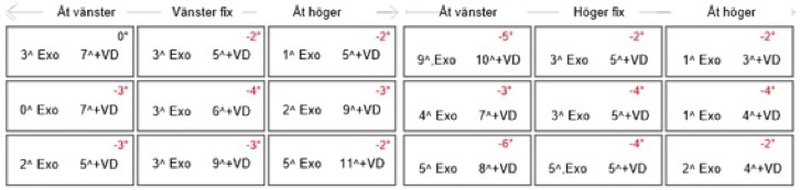
4. Strabismus angle (horizontal and vertical) in the nine cardinal positions of gaze. The angle is given in prism dioptres for fixing with the right eye and fixing with the left eye.
PRODUCT SHEET